Angular deformities of the knee are common during childhood and usually are variations in the normal growth pattern. Angular deformity of the knee is a part of normal growth and development during early childhood. Physiologic angular deformities vary with age as:
- During first year: Lateral bowing of tibia
- During second year: Bow legs (knees and tibia)
- Between 3-4 years: Knock Knees
The condition usually becomes more evident when the child is 2 to 3 years old and normally corrects itself by the time a child is 7 or 8 years old. However, if the condition is not corrected it could be a sign of an underlying disease that requires treatment.
A perfectly aligned knee has its load-bearing axis on a line that runs through the hip, knee and ankle. Based on the inward/ outward inclination of the head of tibia/fibula; knee angular deformities are classified as:
- Genu valgum (knock-kneed): Head of tibia/fibula (not the joint itself), is inclined away from the midline of the body
- Genu varum (bow-legged): Head of tibia/ fibula is inclined toward the midline of the body
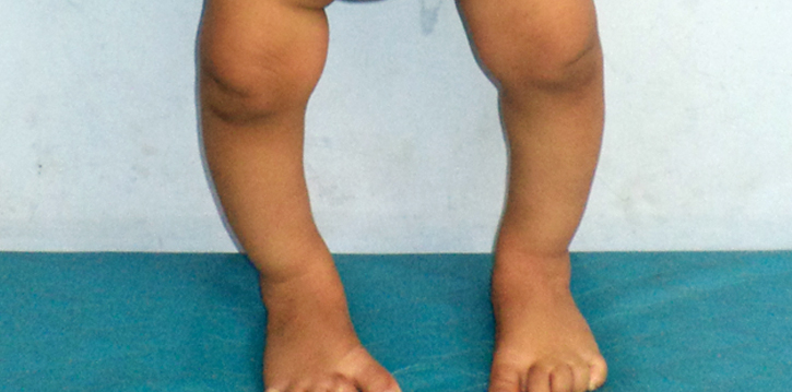
Genu Varum (bowed legs)
Bowed legs are very common in toddlers. If a child has bowlegs, one or both legs curve outwards. When your child stands there is a distinct space between the lower legs and knees. Bowed legs are rarely seen in adolescents. In most of the cases, children with bowed legs are significantly overweight.
The common causes of bowed legs include:
- Physiologic Genu Varum: Most childrenbelow the age of 2, showbowing of the legs as a part of normal physiological process. Normally the bowing will correct by 3 to 4 years of age and the legs may have a normal appearance.
- Blount’s disease: It is a condition in which there is an abnormality of the growth plate at the upper portion of the tibia (shinbone).
- Rickets: It is bone disease that occurs in children due to deficiency of calcium, phosphorus, or vitamin D that are essential for healthy bone growth.
- Trauma
- Infection
- Tumor